
News|Articles|February 1, 2019
- Pharmaceutical Technology-02-01-2019
- Volume 2019 Supplement
- Issue 1
Emerging Therapies Test Existing Bioanalytical Methods
Drug developers must understand the complex bioanalytical assays for cell- and gene-therapy drug development programs and ensure that partners have the specialized expertise needed for complex therapeutic classes.
Advertisement
With many gene and cell-therapy drugs in development, there is an increased need for scientific expertise to develop new bioanalytical technologies as well as use existing platforms such as quantitative polymerase chain reaction (qPCR) and flow cytometry within the regulated bioanalytical space. Current FDA guidance provides little direction on how to approach assay development and validation when using these emerging technologies, nor is there guidance for the application of more familiar technologies to gene-therapy applications. It is important to understand the complex assays needed for these programs and work with partners that provide expertise to ultimately expedite these highly complex therapeutic classes.
Specialty assays used to understand the distribution, function, efficacy, and immunogenicity of gene- and cell-therapy drugs-including cell-based neutralizing assays, qPCR, flow cytometry and enzyme-linked immunosorbent assays (ELISA)-are discussed, as well as recommended strategies for development, validation, and implementation of assays for sample analysis from regulated non-clinical and clinical studies.
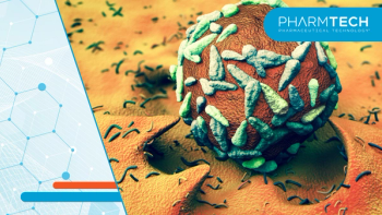
Cellular and gene therapies are not new to the medical establishment. Cellular therapy, the transfer of live cells with a specific function to a patient to treat/cure a disease, was first successfully used in humans in the early 19th century in the form of blood transfusion. Bone marrow transplants, a similar form of treatment, have become relatively common since the initial reported use in 1968.
Gene therapy, the transfer of genetic material into specific cells to modulate gene expression or produce a new/modified protein, was first tested in a clinical trial in 1990 (1). Researchers at the National Institute of Health’s Clinical Center treated two children with adenosine deaminase deficiency using the patient’s own white blood cells that had been harvested, had normal genes inserted, and were reinjected into the patient.
Since 1990, due to improved understanding of the human genome, proteome, and immune system along with improved drug delivery processes, the safety, efficacy, and applicability of cellular and gene therapies seem to be truly coming of age.
Both types of therapy have been shown to cure or mitigate the underlying cause of genetic and acquired diseases by replacing missing proteins or cells, modulating expression of specific proteins, and/or selectively killing tumor cells. Challenges remain for development of these therapies, particularly in the bioanalytical method approach required. Questions include the following:
- How much of a dosed gene therapy is present in circulation, in the target tissue, and how long does it stay there?
- Is the target protein expression changed per the drug mechanism?
- Is the vector genome released (shed) from the patient, allowing for potential transmission to other humans or the environment?
- For cell therapies, do the transferred cells survive and do they express the appropriate markers?
- Do the cells expand in vivo?
The assays used to measure these characteristics are often not traditional liquid chromatography–mass spectrometry (LC–MS) or ELISA used to quantify small- and large-molecule therapeutics and biomarkers. The May 2018 FDA Bioanalytical Method Validation (BMV) Guidance for Industry (2) provides general guidelines for all bioanalytical assays but lacks any specifics on assays that use flow cytometry, PCR, protein expression/activity, or other techniques. This leaves investigators asking questions about how to demonstrate the key bioanalytical attributes specified in the guidance, namely demonstrating that the method measures the intended analyte, evaluating assay variability, the range in measurements that provide reliable data, and the effect of sample collection, handling, and storage on the reliability of data.
Gene therapy assays
For gene therapy programs, one of the first considerations is the biodistribution of the dosed vector and genetic material (3,4). The biodistribution assay will indicate whether the target tissue is successfully transduced by the vector and, if so, how much is present within the tissue. The assay will also determine whether other tissues beyond the specified target are affected; off target transduction will result in toxicity. It is especially important to assess whether the vector reaches the gonads and, if present, whether it is in the germ cells that can result in transmission to the next generation. Sampling from biodistribution studies should be performed at multiple time points and should include time of expected peak concentrations along with multiple late time points to determine both distribution/presence and clearance of the dosed gene therapy. Both the vector and transgene should be evaluated.
Quantitative PCR is typically used for the biodistribution assay because of sensitivity requirements. Assay validation includes evaluation of intra- and inter-assay precision and accuracy, specificity and selectivity, limit of detection, and stability. Expression of the gene therapy product should also be considered, to help determine whether the transgene protein is expressed or if the target protein expression is altered per the dosed gene therapy. The expression may be assessed via a standard ELISA; the current 2018 FDA BMV guidance (2) provides specific assay parameters to validate. These parameters include intra- and inter-assay precision and accuracy, dilutional linearity/integrity, selectivity, and stability, with acceptance criteria of ± 20% bias and ≤ 20% coefficient of variation applied (25% at the lower and upper limits of quantification). In some instances, the activity of the protein may be beneficial to assess, and similar validation parameters and acceptance criteria are generally straight-forward to apply.
Another important assay for gene therapy programs is immunogenicity both to the viral vector and to the product of the transgene. Most of the vectors utilized are likely to have modifications that will trigger the host to recognize it as “foreign” and elicit an immune response that may change the clearance of the vector prior to proper transduction or produce neutralizing effects. In addition, many of the viral vectors may react with antibodies already present in animals and humans. To prevent these pre-existing antibodies from interfering with the program, it is recommended to pre-screen the sample population, and often individuals with high titers of antibodies are excluded from the program.
The assays used to assess immunogenicity are standard ELISAs, cell-based assays are preferred to determine neutralizing effect. The 2016 draft FDA immunogenicity guidance (5) provides the necessary assay parameters to be evaluated, including cut point determination, intra- and inter-assay precision, sensitivity, drug tolerance, specificity, and selectivity. At a minimum, the screening and confirmatory assays should be performed, and when needed, the anti-drug antibodies can be further characterized by performing titer and neutralization assays.
If the delivery mechanism is a viral vector, to determine whether the vector genome was released from the patient resulting in the possibility of horizontal transmission, then viral shedding (release of the vector genome from the patient) should be evaluated to determine the possibility of transmission to other individuals and to the environment. A quantitative approach is recommended, and therefore, qPCR is typically used. Matrices such as urine, saliva, and nasal swabs are tested to determine whether horizontal transmission of the viral vectors occurred. A guidance for this assessment was released in 2015 by FDA (6).
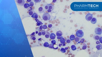
Cell-therapy assays
Cell-therapy products have evolved from blood, bone marrow, and stem cells to more complex engineered autologous or allogenic immune cells to treat cancer. Cell-therapy products that meet the definition of “biologic product” require complex analytical strategies and unconventional bioanalytical methods to determine distribution and persistence of the product. In the case of chimeric antigen receptor T (CAR-T) cells, the construct and viral vector used for transduction are key in determining the design and type of assays required. The bioanalytical methods for these types of programs typically require flow cytometry, qPCR, and immunogenicity assessments.
The first consideration is the development of reagents to be used as controls within the assays. The time upfront in production and characterization of high-quality reagents will result in reduced cost and timelines for the program. Experiments to develop the methods should be carefully thought through to ensure all parameters are evaluated prior to proceeding to validation. High sensitivity methods are required especially when evaluating the expansion and persistence of the CAR-T cells. When considering the development of flow cytometry methods to detect expression of the CAR, optimal antibody and fluorochrome combinations are determined, antibodies are titrated to maximize sensitivity and specificity, and appropriate controls are incorporated (i.e., fluorescence minus ones, experimental procedures for lysing, washing, fixation, intra- and inter-assay precision, limit of detection, inter-instrument assessments, and stability). qPCR method development and validation is similar to that described previously for gene therapy programs. Immunogenicity assay development and validation will follow the recommendations in FDA’s 2016 draft guidance (5); however, one consideration is the detection of antibodies to multiple sequences within the CAR construct that likely will be considered foreign to the recipient’s immune system (i.e., anti-viral protein response).
Platforms for gene- and cell-therapy programs
The choice of platform is primarily driven by and unique to the bioanalytical needs of the gene- and cell-therapy product under development. Specificity, selectivity, and sensitivity requirements also weigh heavily into the final decision. Irrespective of the platform, the choice of reagents has a significant impact on specificity and sensitivity, and therefore time and effort are well-spent in identifying and securing a sufficient and well-characterized supply of reagents to support the lifecycle of all bioanalytical methods.
qPCR is used to quantify a viral genome in serum, tissue, or body fluids for the purpose of pharmacokinetics (PK), biodistribution, or vector shedding evaluations (3,4,6). It is also used to measure the expression levels of transgene- expressed mRNA in target tissue for gene therapy programs. PCR is a widely used platform; however, there is a lack of specific regulatory guidance and industry standards for development and validation. The Minimum Information for Publication of Quantitative Real-Time PCR Experiments (MIQE) guidelines published in 2009 (7) were designed to standardize nomenclature, design, and interpretation of qPCR experiments, and provide a valuable reference in the absence of other publications.
Flow cytometry platforms can measure multiple parameters within a single analysis, and it is a powerful tool for cell-therapy programs. The technology allows for real-time monitoring of the cell therapy in vivo and can support the qPCR data for PK. Flow panels can be designed to evaluate the expression and persistence of the construct over time but can also provide insight into the immune activation status by the addition of antibodies to known markers on lymphocytes. Stability of samples need to be taken into consideration when performing flow for cell-based therapies. To maintain sample integrity, in most instances, samples need to be analyzed within 24–48 hours, which can lead to logistical challenges. The European Bioanalysis Forum released a white paper in 2017 (8) with the intent to provide practice guidance on the use of flow cytometry for regulated studies that support drug development programs. The paper addresses the use of flow cytometry from a bioanalytical perspective and addresses additional parameters not discussed in previous publications.
In genome-editing applications, newer, rapid-sequencing platforms such as next-generation sequencing (NGS) offer techniques to monitor both the efficacy of editing as well as potential off-target gene edits. These technologies are challenging to validate and therefore are typically performed in a non-good laboratory practice fashion. There currently are no regulatory guidance documents that govern this body of work; however, as NGS is used in clinical laboratories for diagnostic purposes, published guidelines (9) governing that work may be leveraged where and when appropriate.
Tried and true platforms such as ELISA maintain a presence in the gene- and cell-therapy bioanalytical space due to relatively low cost, ease of use, and solid performance. ELISA assays can be applied to the detection of anti-capsid or anti-transgene antibodies, and to detection of antibody responses to other components of the gene or cell therapy that may possess immunogenic potential. A requirement for enhanced sensitivity may benefit from newer technologies such as electrochemiluminescence or resonance-based platforms. These newer platforms also offer the advantage of being species-agnostic and therefore can be used throughout the nonclinical and clinical continuum. FDA, along with other global regulatory bodies, have published specific guidance documents for the development, validation, and assessment of anti-drug antibodies (5). Effort should be made to adhere to these guidance documents in the assessment of immunogenicity to gene and cell-therapy elements.
In some situations, the nature of the therapy, molecule, disease, or patient may dictate the need to derive a fit-for-purpose approach to immunogenicity assessment. Cell-based assays provide an essential platform for the detection of neutralizing antibodies. Cells that are permissive to transduction by the viral vector are exploited for this purpose in gene therapy programs. To-date, the design, execution, and reporting of data from these assays has not been standardized, making comparison across studies challenging.
Commercial ELISA kits can be used for the quantification of the newly-expressed protein resulting from gene therapy. In this setting, the measurement of expressed protein is considered a biomarker, and the assay should be validated to the requirements specified in FDA’s May 2018 bioanalytical guidance (2).
More recent technologies such as Simoa (Quanterix) and SMC Errena (Singulex) instruments promise ultra-high sensitivity, which is an advantage when trying to quantify the typically very low levels of protein. Recently, the market has seen an increase in highly sensitive commercial ELISA kits and chemiluminescent and fluorescent substrates that also enhance sensitivity. Other platforms can be applied to the functional assessment of expressed proteins; examples include clotting time for expressed coagulation factors or substrate cleavage by expressed enzymes. FDA released guidance on biomarker evidentiary framework (10) that describes a recommended approach for analytical biomarker assays.
With the newer generation and more complex gene and cell-therapy products in development, it is important to seek scientific expertise to plan and design what is needed to develop and validate the bioanalytical methods required to achieve an overall successful program.
References
1. R.M. Blaese et. al., Science. 270 (5235), 475–480 (1995).
2. FDA, Guidance for Industry: Bioanalytical Method Validation (Rockville, MD, May 2018).
3. FDA, Guidance for Industry: Gene Therapy Clinical Trials-Observing Subjects for Delayed Adverse Events (Rockville, MD, November 2006).
4. FDA, Guidance for Industry: Preclinical Assessment of Investigational Cellular and Gene Therapy Products (Rockville, MD, November 2013).
5. FDA, Draft Guidance for Industry: Assay Development and Validation for Immunogenicity Testing of Therapeutic Protein Products (Rockville, MD, April 2016).
6. FDA, Guidance for Industry: Design and Analysis of Shedding Studies for Virus of Bacteria-Based Gene Therapy and Oncolytic Products (Rockville, MD, August 2015).
7. S.A. Bustin et. al., Clin. Chem. 55(4), 611–622 (2009).
8. B.V. Der Strate, et. al., Bioanalysis. 9(16),1253-64 (2017).
9. R. Somak, et. al., J. Mol. Diag. 20(1), 4-27 (2018).
10. FDA, Guidance for Industry and FDA Staff: Biomarker Qualification: Evidentiary Framework (Rockville, MD, December 2018).
About the authors
Christina Satterwhite, PhD, is senior director, global laboratory sciences; Jessica St. Charles is director, laboratory sciences; Valerie Theobald is director, immunoanalytical services; and Jenifer Vija is scientific director, bioanalysis, all at Charles River Laboratories.
Article Details
Pharmaceutical Technology
Supplement: Partnering for Bio/Pharma Success 2019
February 2019
Pages: s24–s26, s31
Citation
When referring to this article, please cite it as C. Satterwhite, et.al., "Emerging Therapies Test Existing Bioanalytical Methods," Pharmaceutical Technology's Partnering for Bio/Pharma Success Supplement (February 2019).
Articles in this issue
over 7 years ago
Analysis of Sub-Visible Particlesover 7 years ago
Utilizing Analytical Services for Success in Innovationover 7 years ago
Best Practices for Immunogenicity Testing of Biologicsover 7 years ago
Determining Drug Stabilityover 7 years ago
CMO Expansions and Investmentsover 7 years ago
Ensuring Quality Control in Vendor Relationshipsover 7 years ago
A Systematic Approach to Tech Transfer and Scale-UpAdvertisement
Related Content
Advertisement

Advertisement
Advertisement