
|Articles|March 2, 2008
- Pharmaceutical Technology-03-02-2008
- Volume 32
- Issue 3
Raw-Material Authentication Using a Handheld Raman Spectrometer
Author(s)Robert L. Green, Christopher D. Brown
Using a handheld Raman spectrometer, the authors developed methods for 28 commonly used excipients and active ingredients.
Advertisement
Pharmaceutical manufacturing facilities are moving toward 100% inspection of incoming raw materials to confirm the content of each container is verifiable at the molecular level. Current practice requires incoming raw-material containers to be opened and samples to be extracted. The materials are then typically transported to a laboratory for chemical analysis, a process that may take several days or longer, during which time the material is unavailable for production. Technologies used for laboratory identification tests include high-pressure liquid chromatography (HPLC), near-infrared (NIR) and mid-infrared (mid-IR) spectroscopies, and other wet chemical methods (1–6). Among these methodologies, Raman spectroscopy has proven efficient and effective for a wide range of pharmaceutical applications, including identity testing of pharmaceutical raw materials, in-process analysis, and authentication of final dosage forms (7–9).
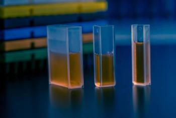
Renewed enthusiasm for Raman technology has been spurred on by numerous technological advances. Raman scattering was historically very difficult to detect because the scattering phenomenon is very weak. NIR lasers, charge-coupled device (CCD) detectors, and Rayleigh rejection filters have made Raman spectroscopy a practical laboratory analysis technique by increasing the sensitivity and decreasing nuisance signal contributions from fluorescence. Furthermore, modern Raman instrumentation is faster, more robust, and less expensive than previous systems. In addition, advances in component miniaturization have led to the availability of portable, handheld Raman systems (see Figure 1).
Figure 1 (ALL IMAGES ARE COURTESY OF THE AUTHORS)
Operational attributes are important to take into account when selecting a measurement technology for a research or manufacturing environment. In the context of material identification, portable solutions (e.g., the handheld Raman instrumentation used in this study) can quickly verify material identity at the point of need. Furthermore, Raman spectra can be acquired through transparent packaging materials such as the plastic bags commonly used to line drums containing incoming raw materials (10, 11). This means of testing reduces the time between receipt and availability to the production line, minimizes handling and, for materials packaged in transparent media, eliminates the risk of contamination posed when the packaging seal is breached. NIR measurements also can be acquired through transparent packaging; however, special care must be taken when making NIR measurements through plastic containers because subtle variations from one container to another can manifest themselves quite markedly in the spectra. In contrast to Raman and NIR, mid-IR techniques require direct contact with the material and are not conducive to measurement through packaging.
In terms of analytical characteristics, Raman spectroscopy is particularly effective for identity testing because of its high degree of selectivity (12). Every chemical compound with covalent bonds produces its own characteristic pattern of Raman shifts, which can be used to chemically fingerprint and therefore identify the compound. Raman spectra for acetylsalicylic acid and acetaminophen are shown in Figure 2, illustrating the excellent selectivity provided by distinct, well-defined peaks.
Figure 2
Some challenges with using other types of spectroscopy, notably NIR, have been method development and method transfer between instruments. As Figure 2 shows, Raman spectra have distinct peaks that are characteristic of the material producing the spectrum. Therefore, like Fourier transform infrared (FTIR) spectroscopy, the intrinsic molecular selectivity means that spectral differences between materials are extremely pronounced relative to nuisance factors. This article describes how Raman methods generated from measurements on one unit can be easily transferred to another, simply by transferring the reference library files.
NIR spectra are less distinct, with broader peaks that result in poorer selectivity, and sometimes require computationally intensive methods to detect differences. As a result, the creation, transfer, and maintenance of NIR methods often require expert oversight and intervention. Variability among various NIR units' optics and other components is sometimes of the same order as the differences among samples tested. As a result, in transferring an NIR method to a different instrument, additional reference spectra and/or tuning of method parameters may be required.
When making an identity assessment based on spectral data, the unknown measurement is examined in relation to one or more reference spectra. A common approach for spectral comparison is to calculate the wavelength correlation, which is equivalent to measuring the cosine of the angle between the two spectra. The resulting correlation coefficient, r, is 1 when the two spectra are in perfect correspondence and 0 when they are orthogonal. Although useful for quick similarity assessments, the correlation coefficient is not particularly sensitive to discrepancies between the two spectra of interest. More problematic, a correlation coefficient other than 0 or 1 has no direct interpretation in the context of spectral identity testing because a transparent interpretation as a test statistic only holds when dealing with random normal variates, which is clearly not the case for FTIR, Raman, or NIR spectra. Despite these difficulties, there is regulatory guidance on selecting a correlation threshold that states, "Unless otherwise justified, a [correlation] threshold below 0.95 is not acceptable..."(13). Arbitrary designation of a correlation threshold in this manner can be perilous because it is unsupported by either basic statistics or demonstration, a point that has also been made by other researchers (14).
Figure 3
Figure 3 shows a Raman reference spectrum for pure glycerin and a Raman spectrum measured for an "unknown" substance, in this case glycerin contaminated with 20% diethylene glycol by volume. Contamination of glycerin with diethylene glycol is a problem of current interest, as evidenced by several news reports and a recent FDA guidance (15). In spite of clear differences in the highlighted regions of the spectra in Figure 5, there is a very favorable correlation coefficient (0.96), which indicates that this material would pass as acceptable unless a higher than typical correlation threshold were applied.
Figure 4
An alternate approach to wavelength correlation used by the handheld units for this study is to evaluate whether the measured spectrum lies within the multivariate domain of the reference spectrum (or spectra), which is defined by the uncertainty characteristics of each measurement, including exposure settings, instrument and environment properties (e.g., temperature, dark current, ambient lighting), and the optical properties of the sample itself. When comparing spectra in this manner, the analyst looks for spectral features that contradict the reference spectrum given the uncertainty of the measurement, rather than how well the bulk spectrum matches. For identity testing, the critical question is whether the measured spectrum can be considered consistent with the reference spectrum given the multivariate uncertainty of the measurement conditions. Like most statistical tests, the analysis is distilled to a p-value, in this case the probability that the observed differences between the test and reference spectra simply arose by chance given the uncertainty of the measurement. Higher p-values indicate that any differences are not large relative to the uncertainty of the measurement. In such cases, the measured spectrum is deemed consistent with the reference spectrum, and the instrument declares "pass." If the p-value is too low (below 0.05 as the device default), then it suggests that discrepancies between the measured and reference spectrum were unlikely to arise from the uncertainty in the measurement alone, and the device declares "fail." The system logic just described is illustrated in Figure 4. The earlier example for the spectra in Figure 3 resulted in a p-value of 3.2 × 10⁻³, which indicates that there is a discrepancy.
Figure 5
Experiment
Handheld Raman material identity verification systems were used in this work (TruScan, Ahura Scientific, Wilmington, MA). Six units manufactured at different times from different runs of parts were used in this study (details provided in subsequent paragraphs in this article). This instrumentation uses a 785-nm NIR external cavity-stabilized, cooled laser as a light source. The laser has a maximum power consumption of 1 W, a maximum output of 400 mW, and typically operates at 300 mW output. A single-dispersive spectrometer, cooled CCD, and dielectric notch filters for Rayleigh rejection make up the rest of the instrument hardware. The device also has an integrated barcode reader for capturing sample lot–batch–other identifiers and selecting methods. Detailed specifications are listed in Table I.
Table I: TruScan instrument specifications.
Three TruScan devices (hereafter referred to as the "test devices") were tested with each of the 32 common pharmaceutical materials listed in the sidebar "Materials tested" (Sigma-Aldrich, St. Louis, MO) to evaluate the applicability of the systems for incoming inspection.
Materials tested
To create methods, reference spectra first had to be acquired. The reference spectra for the materials were taken on three TruScan devices (hereafter referred to as "reference devices") using identical data collection software as the test devices described above. In the context of TruScan, methods are analytical tasks based on stored reference spectra that the instrument's software executes to determine whether a material's identity can be verified. For each material, a single reference spectrum was acquired by one of the three reference devices.
To collect each reference spectrum, a sample of the material was placed in a borosilicate glass vial (VWR, West Chester, PA) and the data were collected through the wall of the vial. The laser aperture of the instrument was placed at the appropriate distance from the vial using either the nose-cone or vial-holder attachment for correct spacing. For most materials, the acquisition process was initiated and simply allowed to continue until terminated automatically by the unit's library acquisition software. In contrast, cellulose, dextrin, trimagnesium phosphate, zinc sulfate, and calcium sulfate are slightly fluorescent, so special care had to be taken to avoid photobleaching the sample during reference measurement. This can be achieved by terminating the reference scan before the software automatically discontinues measurement or by periodically moving the sample throughout data collection. Finally, while reference spectra were collected, it was determined that colloidal silica, talc, sodium carboxy methyl cellulose, and hydroxy propyl methyl cellulose did not provide an adequate signal to be measured in a practical period of time for handheld deployment. In particular, colloidal silica had a very weak Raman signal, and the other three materials were too fluorescent to allow reliable determinations to be made.
Methods were created from the reference spectra by the associated web-based software utility and then loaded into the test instruments. To prepare test samples to challenge the methods, samples of approximately 2 g each of the 28 remaining materials were sealed in 2-m-thick polyethylene bags in an effort to emulate expected-use scenarios (measurement through plastic bags) for incoming material inspection. Three measurements of each sample were made, one measurement with each of the three test devices. Each test measurement was made using the automatic (or "auto") mode, where the unit's software controls data acquisition parameters to achieve the necessary spectral signal-to-noise ratio (SNR) in the shortest measurement time possible. The auto-mode measurements used when running a method compensate for differences in operator positioning, stray-light, lot-to-lot sample fluorescence, and so forth. Each "unknown" measured spectrum (three for each material) was evaluated against each method for the test materials using the probability based approach described previously, resulting in a p-value for each unknown-method pair.
Results and discussion
When authenticating material, the user selects the appropriate method from a menu on the instrument's display and enters the material identification number with the barcode reader or keypad. The operator directs the unit's laser aperture toward the sample and initiates the measurement. Once data collection begins, the instrument optimizes settings including exposure time, number of accumulations, and laser power to achieve acceptable SNR as quickly as possible. Table II shows the average measurement times required for each material. As demonstrated by the information in Table II, the average measurement times vary considerably depending on the characteristics of the material (Raman cross section, etc.). Specifically, the times range from 1 s to more than 6 min; however, the majority of materials were measured in less than 1 min. The individual measurement times (not shown) for the three units differ to some degree, as well, primarily because of environmental conditions during the measurement (sample positioning, ambient light, etc.).
Table II: Measurement time averages (rounded to nearest second).
As indicated in the experimental section, a p-value for each unknown-method pair was generated for measurements across all three test devices. The p-values, which indicate whether the measured spectrum is statistically different from the reference spectrum, were examined individually and were averaged for presentation purposes. Figure 6 shows data for each material– method pair. Note that the values shown range from p <10–15 to p >0.1. In normal application of the instrument, the result is a simple pass–fail determination. The unit's default threshold for "pass" is p ≥0.05. This indicates that the measured spectrum is statistically consistent with the reference spectrum given the uncertainty of the measurement.
Figure 6
The diagonal elements of Figure 6 represent cases in which the material was tested against the corresponding method, whereas the off-diagonal entries represent materials being tested by the "wrong" method. The dark-green squares on the diagonal show where the measured spectrum matched the corresponding reference spectrum with a p-value >0.1, which indicate that these materials easily pass as being consistent with the method reference spectrum. Two materials, ethyl cellulose and hydroxypropyl cellulose, resulted in average p-values in the range of 0.01 to 0.1 when tested against themselves. As also shown in Figure 6, examination of individual results from each of the three instruments revealed that these two samples, numbers 10 and 11 (indicated by daggers), were the only ones that resulted in differences in pass–fail decisions among the three test instruments. For ethyl cellulose, the p-values were 0.03, 0.06, and 0.04, and for hydroxypropyl cellulose, the p-values were 0.02, 0.05, and 0.11 across the three test units, respectively. Thus, both materials were very close to the borderline threshold of 0.05 and either just passed or just failed.
As shown in Table II, ethyl cellulose and hydroxypropyl cellulose also correspond to the longest required measurement times, 284 s and 390 s, respectively. Further inspection of the spectral data (not shown) revealed subtle features in the unknown measurements that were not present in the reference measurements. To determine whether the polyethylene containers could be responsible, an auto-mode spectrum for each of these two materials was acquired through borosilicate glass vials. These spectra were consistent with the reference measurements and did not contain any extra features. In addition, a spectrum of polyethylene was acquired by folding an empty polyethylene bag over on itself several times to produce a sample of sufficient thickness for measurement. The resulting spectrum (not shown) contained bands in the spectral regions where the extra peaks in the unknown were found. Thus, despite the ability of Raman to sample through packaging, the extremely weak signal and long measurement times for these two cellulose materials resulted in conditions favorable to allow the plastic to subtly interfere with the measurement. It is likely that measurements could be successfully made through glass, but this was not attempted because the required measurement time may not be considered practical for routine field use.
Examination of the off-diagonal elements in Figure 6 confirms the excellent selectivity of the technology as evidenced by the overwhelming majority of off-diagonal elements with p < 10–15. The only area in the table where there is a lack of acceptable selectivity is for the alkali metal stearate materials. Stearic acid is differentiable from both calcium and magnesium stearate (p < 0.01); however, calcium stearate and magnesium stearate cannot be readily differentiated from one another. Whereas many materials differing only in their cation can be readily differentiated with the handheld Raman system (e.g., in the study, sodium and potassium bicarbonate, calcium, and zinc sulfate), calcium stearate and magnesium stearate are simply too similar from a spectroscopic standpoint. This is likely a result of the change in cation not having an appreciable long-range influence on the relatively large anion (stearate) from which the Raman signal is actually generated.
Conclusion
Handheld Raman spectroscopy is an excellent alternative to traditional incoming raw-material inspection by high-pressure liquid chromatography, wet chemical methods, and NIR and mid-IR spectroscopy. The technology has excellent specificity, which, coupled with intelligent on-board algorithms, reduce the time and effort required to develop and validate methods. Furthermore, methods can be successfully loaded onto different handheld Raman instruments to produce consistent data and material identification on the multiple instruments, without loading additional spectra or performing other customization.
In addition to its analytical characteristics, today's handheld Raman solutions are environmentally robust and can be used by expert spectroscopists as well as operations-based personnel. This is in contrast to Raman instruments of the past, which were bulky, slow, expensive, and delicate. Based on the study presented in this article of common pharmaceutical materials, the handheld Raman spectrometer offers an attractive option for 100% inspection of most incoming raw material in pharmaceutical manufacturing facilities.
Acknowledgment
Dr. Wayne Jalenak is gratefully acknowledged for useful discussions and for preparation of the glycerin samples presented in the introduction.
Robert L. Green* is a research scientist and Christopher D. Brown is a director of system analytics and applications, both at Ahura Scientific, Inc., 46 Jonspin Road, Wilmington, MA 01887, tel. 978.657.5555, fax 978.657.5921,
*To whom all correspondence should be addressed.
Submitted: July 26, 2007. Accepted: Sept. 13, 2007.
References
1. H.T. Rasmussen et al., "Validation of HPLC Methods in Pharmaceutical Analysis," in Handbook of HPLC in Pharmaceutical Analyses, S. Ahuja and M. Dong, Eds. (Elsevier, San Diego, United States, 2005), pp. 192–216.
2. M.A. Dempster et al.,"A Near-Infrared Reflectance Analysis Method for the Noninvasive Edentification of Film-Coated and Non–Film Coated, Blister Packed Tablets," Anal. Chim. Acta. 310, 43–51 (1995).
3. M. Blanco et al., "Near-Infrared Spectroscopy in the Pharmaceutical Industry," Analyst 123, 135R–150R (1998).
4. P.J. Gemperline, L.D. Webber, and F.O. Cox, "Raw Materials Testing Using Soft Independent Modeling Class Analogy Analysis of Near-Infrared Reflectance Spectra," Anal. Chem. 61 (2), 138–144, (1989).
5. J.A. Ryan et al., "Rapid Verification of Identity and Content of Drug Formulations Using Mid-Infrared Spectroscopy," J. Pharm. Biomed. Anal. 9 (4), 303–310 (1991).
6. For example, United States Pharmacopeia-National Formulary and European Pharmacopeia.
7. T. Vankeirsbilck et al., "Applications of Raman Spectroscopy in Pharmaceutical Analysis," TrAC 21 (12), 869–877 (2002).
8. R.L. McCreery et al., "Noninvasive Identification of Materials Inside USP Vials with Raman Spectroscopy and a Raman Spectral Library," J. Pharm. Sci. 87 (1), 1–8 (1998).
9. R. Cantu et al., "A Simple Approach for Developing, Validating, and Transferring an Identification Method for Multidose Pharmaceutical Products using FT-Raman Spectroscopy," Am. Pharm. Review 10 (2), pp. 96–103, 111 (Jan.–Feb. 2007).
10. D.A.C. Compton and S.V. Compton "Examination of Packaged Consumer Goods by Using FT-Raman Spectrometry," Applied Spec. 45 (10), 1587–1589 (1991).
11. S.G.Skoulika and C.A.Geurgio, "Rapid, Noninvasive Quantitative Determination of Acyclovir in Pharmaceutical Solid Dosage Forms Through their Poly(vinyl chloride) Blister Package by Solid-State Fourier Transform Raman Spectroscopy," Applied Spec. 57 (4), 407–412 (2003).
12. R.L. McCreery, "Introduction and Scope," in Raman Spectroscopy for Chemical Analysis (John Wiley & Sons, New York, United States, 2000), pp. 1–14.
13. European Medicines Agency (EMEA), Note for Guidance on the Use of Near Infrared Spectroscopy by the Pharmaceutical Industry and the Data Requirements for New Submissions and Variations (London, UK, 2003).
14. C.T. John and N.C. Pixley "Methodology for NIR Identification of Pharmaceutical Finished Products with Emphasis on Negative Controls and Data Driven Threshold Value Selection," Am. Pharm. Review, 10 (2), 120–124 (Jan–Feb 2007).
15. FDA, Center for Drug Evaluation and Research, Guidance for Industry: Testing of Glycerin for Diethylene Glycol (Rockville, MD, 2007).
Articles in this issue
almost 18 years ago
FDA Seeks Regulatory Flexibilityalmost 18 years ago
Time for a World Tooling Standardalmost 18 years ago
Report from: Indiaalmost 18 years ago
Holding Backalmost 18 years ago
Containers Insulate Companies from Product Lossalmost 18 years ago
Inside USP: Metrology and USP Dissolutionalmost 18 years ago
Pharmaceutical Technology's Equipment and Machinery Trends Surveyalmost 18 years ago
Coming Down the Pike: HDAC inhibitorsNewsletter
Get the essential updates shaping the future of pharma manufacturing and compliance—subscribe today to Pharmaceutical Technology and never miss a breakthrough.
Advertisement
Related Content
Advertisement


Advertisement
Advertisement
Trending on Pharmaceutical Technology
1
Drug Digest: Outsourcing Partnerships Fuel Global Biopharma Discovery and Scale-Up
2
FDA Grants Priority Review For Pfizer’s Marstacimab for Hemophilia A or B
3
Shaping the Future of Microbial Development: From Gene to GMP
4
PharmTech Weekly Roundup - February 6, 2026
5